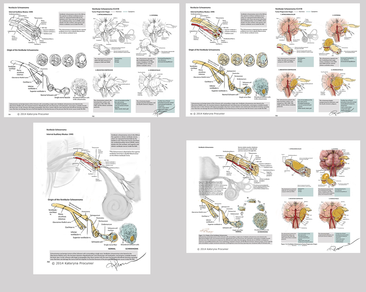
Title: Vestibular Schwannoma
Year: 2014
Awards: AMI Orville Parks Student Best of Show 2014 | AMI Award of Excellence 2014
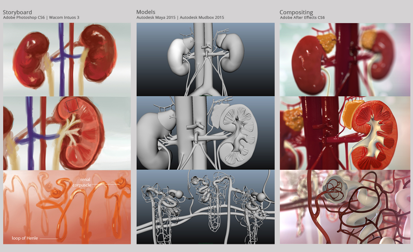
Medium: Adobe Photoshop CS6 | Wacom Intuos 3 | Layout in Adobe Illustrator CS6
Audience: First year medical students
Supervisors: Dr. Linda Wilson-Pauwels | Andrea Gauthier
Content Expert: Dr. Dee Ballyk
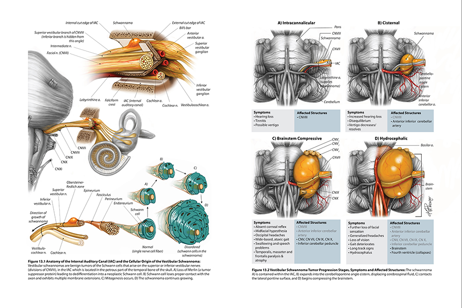
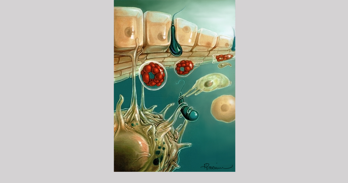
Purpose: This illustration is intended as a supplementation to the content taught to first year medical students about cranial nerves and cranial nerve lesions. The narrow space inside the skull where the cranial nerves exit the brainstem makes it very difficult to learn about their spatial relationship to tumors such as the vestibular schwannoma. I chose an anterior angle of view of the brainstem because I felt that this was the best method to visualize this complex relationship, making it clear as to why the nerves become affected in the order that they do as the disease progresses, and how this relates to the clinical symptoms. The cellular development of the schannoma was also included to show the origin of the disease.
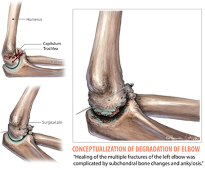
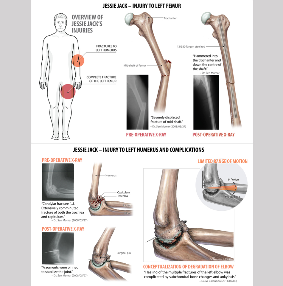
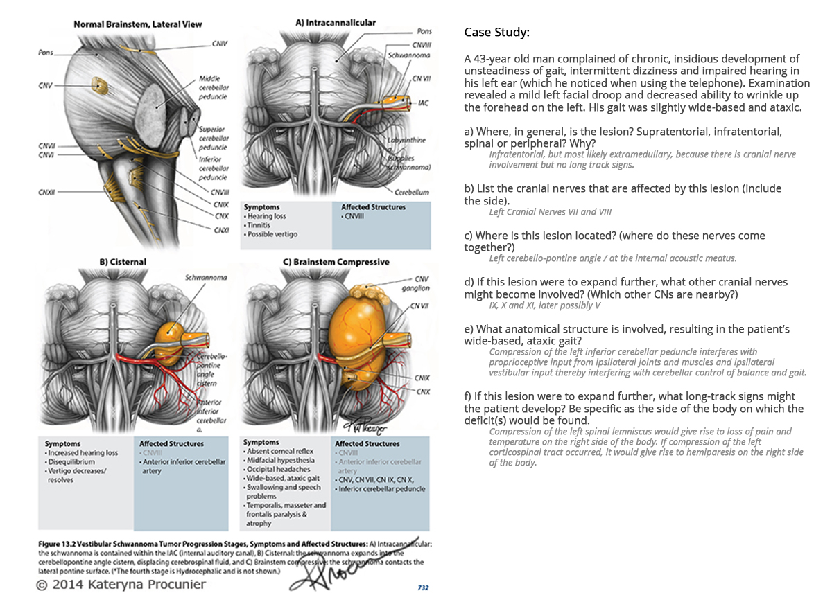
Case Study
A slightly different layout was also created as requested by my content expert, involving a lateral view of the brainstem, and specific for the following case study:
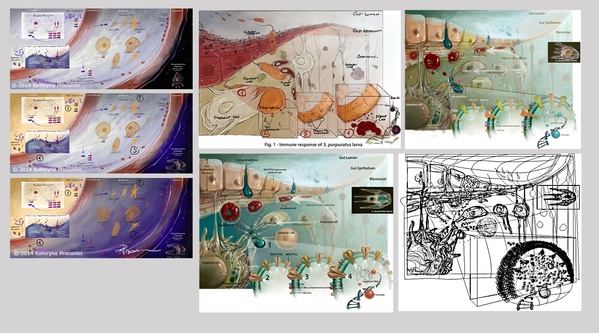
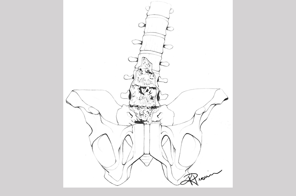
Sketches
References
Brunsteins, D. B. and Ferreri, A. J. (1995). Microsurgical anatomy of arteries related to the internal acoustic meatus. Acta Anatomica, 152(2), 143-150.
Glastonbury, C. M et al. (2002). Imaging Finding of Cochlear Nerve Deficiency. American Journal of Neuroradiology, 23, 635-643.
Hanemann, C. O. (2008). Magic but treatable? Tumours due to loss of Merlin. Brain, 131, 606-615.
Hung, G. et al. (2002). Immunohistochemistry Study of Human Vestibular Nerve Schwannoma Differentiation. Glia, 38, 363-370.
Jackler, R. K. and Brackmann, D. E. (2005). Neurotology (2nd ed.). Philadelphia, PA: Elsevier Mosby.
Kim, H.-S. et al. (1998). Topographical relationship of the facial and vestibulocochlear nerves in the subarachnoid space and the internal auditory canal. American Journal of Neuroradiology, 19, 1155-1161.
Klenke, C. et al. (2013). Clinical and biological behaviour of vestibular schwannomas: signalling cascades involved in vestibular schwannoma resemble molecular and cellular mechanisms of injury-induced Schwann cell differentiation. Head Neck Oncology, 16, 5(2), 20.
Özdoğmuş, Ö. et al. (2004). Connections between the facial, vestibular and cochlear nerve bundles within the internal auditory canal. Journal of Anatomy, 205, 65-75.
Schuenke, M. et al. (2010). Thieme atlas of anatomy: head and neuroanatomy. HongKong: Everbest Printing Ltd.
Sethi, A. et al. (2011). Intraparotid facial nerve neurofibroma: and uncommon neoplasm. International Journal of Morphology, 29(3), 1054-1057.
Snow, J. B., Wackym, P. A., & Ballenger, J. J. (2009). Ballenger’s Otorhinolaryngology: Head and Neck Surgery. USA: PMPH.
Stonecypher, M. S. et al. (2006). Neuregulin Growth Factors and Their ErbB Receptors Form a Potential Signaling Network for Schwannoma Tumorigenesis. Journal of Neuropathology & Experimental Neurology, 65(2), 162-175.
Yamakami, I. et al. (2002). Hypervascular vestibular schwannomas. Surgical Neurology, 57(2), 105-112.
Young, B. et al. (2006). Wheater’s Functional Histology: A Text and Colour Atlas. Elsevier Limited.