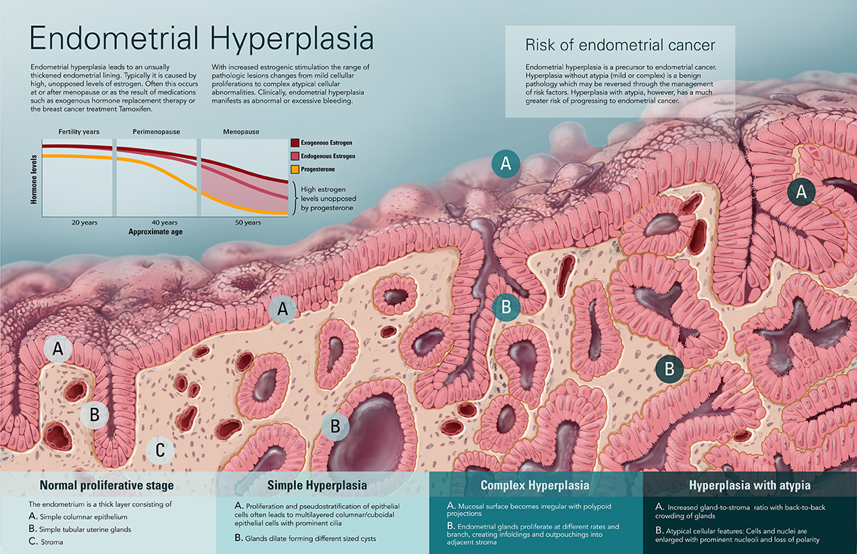
Endometrial Hyperplasia
- Media:
- Adobe Photoshop
- Target Audience:
- Educated lay audience
- Format:
- Supervisor:
- Prof. Dave Mazierski, Biomedical Communications, University of Toronto
- Content advisor:
- Dr. John Wong, Laboratory Medicine and Pathobiology, University of Toronto
- Purpose:
- To illustrate hyperplastic changes in uterine tissue most common among women who are nearing or have reached menopause. Demonstration of etiology, pathogenesis, morphological changes and clinical manifestations of endometrial hyperplasia at various stages of classification.
PROCESS
After researching endometrial hyperplasia and the normal versus pathological histological features of the inner uterine lining, sketches were created in order to visualize the tissue in greater detail.
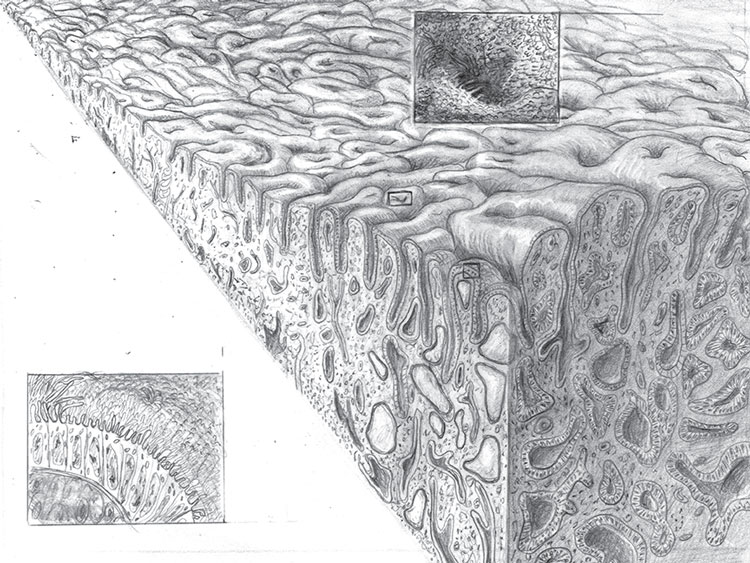
Next, a comprehensive sketch was developed in pencil to illustrate a tissue landscape that aims to depict not only the increase in cell number but also to explain the pre-cancerous disease classifications by illustrating relevant alterations in gland and stromal architecture.
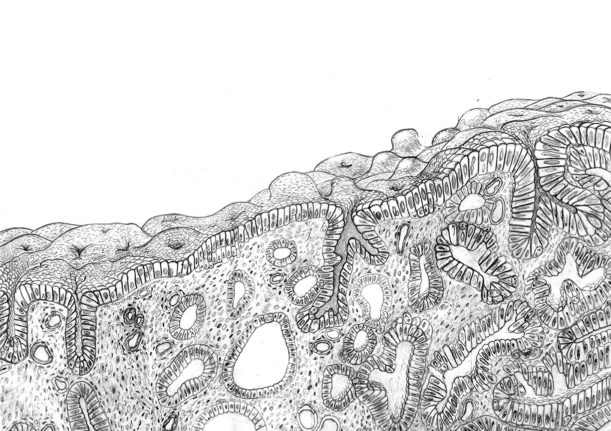
The sketch was then scanned, cleaned and painted in photoshop. Adobe Illustrator was used to layout the illustration and create the graphical and textual information.

References:
Aplin, J.D., T. Asgerally Fazleabas and Linda C. Giudice. 2008. Endometrium : Molecular, cellular, andclinical prespectives. New York, NY, USA: CRC Press.
Color atlas and text of histology /Leslie P. Gartner, James L. Hiatt.
Deligdisch, L., and C. J. Cohen. 1985. Histologic correlates and virulence implications of endometrial carcinoma associated with adenomatous hyperplasia. Cancer56 : 1452-5.
Deligdisch, L., G. Yedwab, A. Persitz, and M. P. David. 1978. Ultrastructural features in normal and hyperplastic postmenopausal endometrium. Acta Obstet Gynecol 57 : 4439-52.
Ferenczy, A. 1994. Anatomy and histology of the uterine corpus.
Endometrial hyperplasia. 2010. Seminars in Diagnostic Pathology 27 (4) (201011): 199-214.
Gusberg, S. B. 1974. Precursors of corpus carcinoma: Estrogens and adenomatous hyperplasia. American Journal of Obstetrics and Gynecology 54 : 905-27.
Marc Hannemann, M., H. Mary Alexander, N. Jane Cope, and N. Acheson. 2007. Endometrialhyperplasia. Obstetrics, Gynaecology and Reproductive Medicine 17 (6): 169-72.
Mills, Anne M., and Teri A. Longacre. 2010. Endometrial hyperplasia. Seminars in Diagnostic Pathology 27 (4) (201011): 199-214.
Sivridis, Efthimios, and Alexandra Giatromanolaki. 2013. Demystifying endometrial hyperplasia. Diagnostic Histopathology 19 (7): 223-30.
Robbins, Stanley L., and Ramzi S. Cotran. 1979. Pathologic basis of disease. Philadelphia: W.B. Saunders Co.