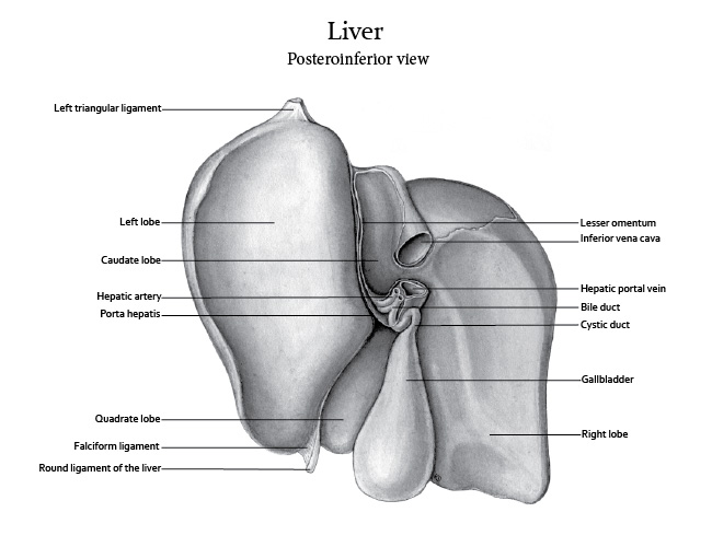
Medical Illustration of the liver
- Media:
- Watercolour
- Target Audience:
- First year medical or pre-medical students
- Format:
- Supervisor:
- Prof. Dave Mazierski, Biomedical Communications, University of Toronto
- Purpose:
- This illustration was created as part of a Human Anatomy and Embryology course and was based on a preserved specimen in Grant's Museum at the University of Toronto.
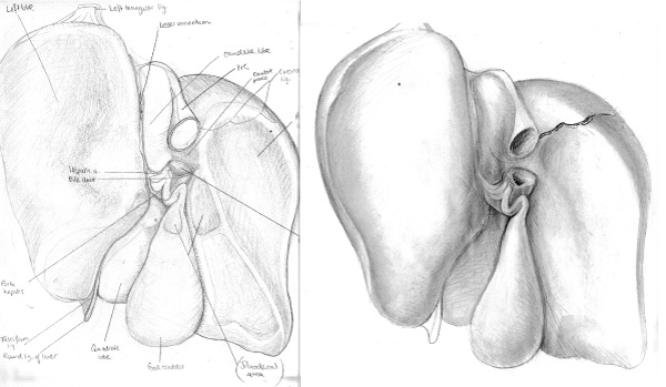
PROCESS WORK
Preliminary pencil sketches identifying the relevant anatomy on the posteroinferior side of the liver. Since the watercolour wash technique is not as forgiving as carbon dust I wanted to make sure I understood the form correctly before starting so I completed a more detailed pencil study of the specimen.
References:
Gray, H., and Williams, P. L. (1989). Gray’s anatomy (37th ed.). Edinburgh: C. Livingstone.